Inter-Chem-Net®

Software Instruction Manual

Version 2000

Introduction

This manual introduces you to the Inter-Chem-Net® (ICN) software at the University of Maine. The Inter-Chem-Net® program allows introductory chemistry students to use sophisticated instrumentation and computer software. The ICN software allows you to:

      1. Collect data using a Ultraviolet-Visible Spectrophotometer
      2. Collect data using an FTIR Spectrophotometer
      3. Analyze data collected with either spectrophotometer
      4. Perform simulations of chemistry labs
      5. Use computerized tutorials to learn chemical concepts

Chemists use sophisticated instrumentation to collect and analyze chemical data and create models of the atomic world. Thus, learning chemistry means understanding it through the perspective of computerized graphs and models.

The Instruments

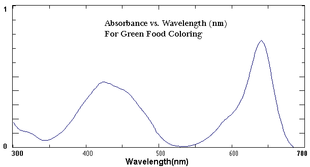
Beckman Ultraviolet-Visible (UV-vis) Spectrophotometer

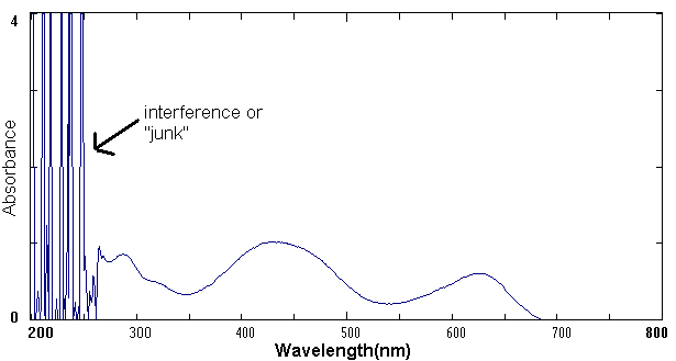
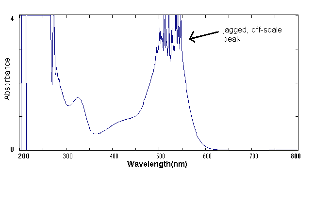
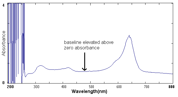
The Inter-Chem-Net® software allows you to use Beckman UV-vis spectrophotometers and Midac FTIR Spectrophotometers. Both instruments are examples of "state of the art" spectrometers used in advanced research and commercial laboratories. The UV-vis Spectrophotometer exposes a chemical solution to ultraviolet and visible light across the electromagnetic spectrum from about 300 to 800 nm. A detector then measures the amount of light absorbed by the solution. The amount of light that reaches the detector is then recorded as a spectrum as shown in the example below. 

Example of a UV-visible Spectrum

Midac FTIR Spectrometer

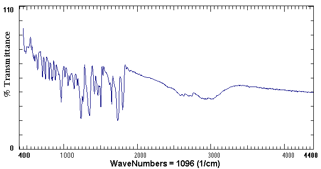
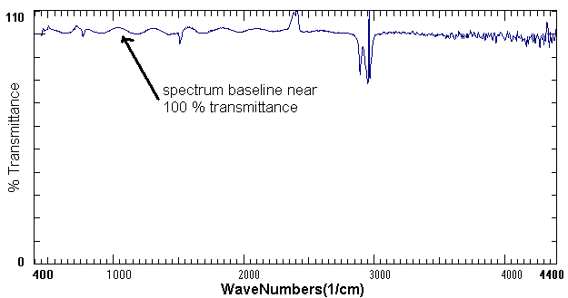
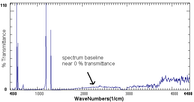
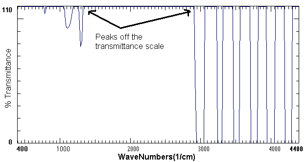
The FTIR or Fourier Transform Infrared Spectrometer exposes a chemical solution to the infrared range of the electromagnetic spectrum. A detector then measures the amount of light transmitted through the solution. The amount of light reached by the detector is then recorded as a spectrum as shown in the example below.

Example of an FTIR Spectrum of Aspirin


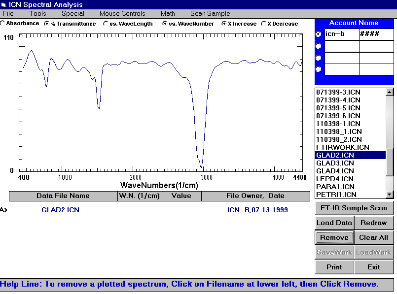
Both types of instruments are accessed through the Inter-Chem-Net® software. Through the Inter-Chem-Net® software, spectra (the plural of spectrum) can be collected quickly and easily on these instruments. There are four UV-vis spectrometers and three FTIR spectrometers in Room 221 Aubert Hall.

Help Resources

For help with Inter-Chem-Net::

    1. consult this manual
    2. choose HELP under the top menu of the ICN software
    3. ask your laboratory TA or another student
    4. ask Bob Kirk (581-1183 or kirk@maine.maine.edu) in Room 230 Aubert Hall.

Where to Access the Inter-Chem-Net® software

The ICN software is available in the Undergraduate Computer Center in Room 421 Aubert Hall. The software is also available through the web address: http://icn2.umeche.maine.edu/icn/ The Inter-Chem-Net® website also contains more valuable information on ICN including the theory of spectroscopy, a tour of ICN, and links to the general chemistry labs.

How to Activate Your ICN Account

Find your 12 digit barcode in your laboratory handbook.

Enter the following information:

If any information is incorrect, choose the ReDo button and correct the information. Choose the Create Account button. Write down your account and password.

How to Use the UV-vis Spectrometer

The UV-vis Spectrometers are located in room 227 Aubert Hall.

    1. Move the Barcode Wand over your barcode from top to bottom OR enter your account name.
    2. The UV lamp should be on. If the UV lamp button in the lower right hand corner reads "UV lamp OFF," click on it so that it reads "UV lamp ON." It takes 30 seconds to warm up.
    3. Fill a cuvette half full of distilled water. Wipe the cuvette with a Kimwipe to remove any oil or fingerprints. Insert the cuvette into the instrument holder with the wavy lines on the cuvette facing the front of the instrument. The light beam needs to pass through the clear sides of the cuvette. Click on the BLANK button. You should hear a click-click sound from the instrument. The blank records what the solvent and cuvette "look like." The instrument then will electronically subtract the cuvette and solvent when it records a spectrum.
    4. Fill a cuvette half full of your chemical sample. Place the cuvette in the instrument holder. Click on the Scan button. The instrument automatically creates a computer file that contains each data set. Repeat for each chemical sample. Write down the filename for every sample. You will need the filename to analyze your data.
    5. Click on the exit when you finish scanning your samples.
    6. Take your samples back to the lab.
    7. Proceed to Room 421 to analyze the experimental spectra.


How to Use the FTIR Spectrometer

The FTIR Spectrometers are located in Room 227 Aubert Hall.

    1. At the prompt, enter your account name.
    2. Place a blank sample card in the holder. Click on the Blank button. The blank records what the sample card "looks like." The instrument then will electronically subtract the sample card when it records a spectrum.
    3. Place your chemical sample card in the FTIR sample holder and click on the Scan Sample button. Record the filename of each sample scan. You will need the filename to analyze your data. Follow the instructions on the bottom of the instrument screen.
    4. Click the Exit button when you finish scanning your samples.
    5. Take your samples back to the lab.
    6. Proceed to Room 421 to analyze the experimental spectra.

How to Analyze Your Data

Go to a computer terminal running the Inter-Chem-Net® program. If your computer is not on yet, turn it on. Enter the login name: PUBLIC and password: PUBLIC. If the ICN program does not start automatically, click on the Inter-Chem-Net® logo. You should now see the ICN Home Page (see front cover of this manual.) Click on Spectra Analysis.

Type in your account name and password. A list of your data files should appear. You can then click on them to view and manipulate the data. For instance, you can change the axis of either the x or y axis scale by highlighting a value and entering a new one. If you click the mouse button and hold it on the screen, a cross bar will appear.

Follow instructions given in lab manual for each particular lab.

Computer Simulations and Tutorials

In addition to the ability to analyze spectra, the Inter-Chem-Net® software offers many tools to help learn the concepts in chemistry. These tools can be used independently of the lab and support the material presented in the lecture portion of the course. These tools include tutorials, simulations, and lots of chemical facts. All of them can be accessed through the main menu by choosing Toolbox. A brief description of each of the tools follows.

References

Periodic Table- displays the Periodic Table with atomic weight, density, melting point, boiling point, ionic radii, electron affinity, ionization energy, heat of atomization, electronegativity, convalent radii, log (cosmic abundance), and number of isotopes. It will also graph any of this periodic information according to atomic number, period, or group.

Electromagnetic spectrum - provides wavelength, frequency, and photon energy for any wavelength in the visible spectrum.

Orbital shapes - provides a graphical view of the atomic orbital shapes from 1s through 4d.

Quantum numbers - shows the quantum numbers corresponding to a given orbital.

Electron configurations - gives the electronic configuration according to the Aufbau principle and spectroscopic notation for any element.

Molecular polarity - provides the polarity of a molecule based on the shape of the molecule and electronegativity of the individual atoms.

Phases of the elements - displays the phases of the elements in the periodic chart at varying temperatures.

Tools

Plotter - creates a plot of any data entered into the spreadsheet.

Equation balancing - a tutorial and practice on balancing equations.

Molecular weight - calculates the formula weight and percent composition for a given formula.

Solution making - calculates the formula weight of a given substance needed to make a given molar solution.

Ionic compounds - predicts the solubility of selected ionic compounds.

Molecular structure - a 3D imager for the structure of molecular structures.

Concepts

Coulomb’s Law - calculates the magnitude of electrical force in Newtons for various charges at a distance of different distances.

Radioactive Decay - demonstrates the rate of decay for four different isotopes: uranium-238, radon-222, hydrogen-3, and carbon-14. It graphs the amount of decay for a given time period.

Atomic Absorption and Emission - simulates the effects of a light source of selected wavelength hitting a hydrogen atom in its ground state.

Bond Energy - Heats of Reaction calculates the heats of reaction for varying bond energies of the reactants and characterizes the reaction as endothermic or exothermic.

Gas Phase Boltzman Distribution - produces a distribution plot of the number of molecules at a particular speed for a selected temperature. It also calculates the fraction of molecules above 1000 m/s.

Liquid Phase Boltzman Distribution - graphs the distribution of molecules at a given energy and shows the percentage of molecules above the energy needed to escape from the surface of the liquid.

Reaction Rates - graphs the concentration of reactants [A] versus time for the conditions you have set and shows an energy diagram of the relative energy levels of the reactants, the transition state, and the products.

Simulations

Limiting Reagents - determines the limiting reagent and amount of product formed for five possible reactions.

Specific Heat - simulates the specific heat by heating blocks of wood, copper, and glass in a Bunsen burner.

Calorimetry - simulates the temperature change in five minutes for a known quantity of a chemical burned in an insulated water bath.

Enthalpy of Dissolution - simulates the energy released or absorbed when a substance is dissolved in water.

Colligative Properties - calculates the changes in boiling point elevation and freezing point depression when a solute is added to water.

Gas Laws - shows how pressure, mass, and temperature affect the volume of a confined gas.

Vapor Pressure - demonstrates the changes in vapor pressure for changing temperatures.

Acids and Bases - shows pH for selected molarities of acids and bases.

pH Titration - simulates an acid/base titration with a corresponding titration curve and equivalence point.

Buffers pH - give the pH for various buffer solutions.

Gibb’s Law - graphs the Gibb’s energy versus the temperature for selected reactions. It also gives the entropy and enthalpy values for each reaction.

Electrolysis - simulates an electrolysis reaction and calculates the time of the reaction, the moles of selected metal deposited on the electrode, and the grams of metal deposited.

Iron Thiocyanate - demonstrates the chemical equilibrium for iron thiocyanate at various concentrations.

Rate Measurement - calculates the reaction rate for the general equation A -à B. It graphs the concentration, [A], versus the elapsed time in minutes.

Data Analysis

Mass Spectroscopy - simulates the mass fragmentation patterns for various molecules.

UV-VIS Spectroscopy - simulates a spectrum for various ionic samples taken at different concentrations.

Electrochemical Cells - simulates a working electrochemical cell.

Spectra library of Common Questions and Mistakes

UV-vis Spectrum #1

UV-vis Problem #1: interference or "junk" in the 200-300 nm range.

UV-vis Solution #1: The plastic cuvettes produce interference in the 200-300nm range. You will see this interference in any spectra produced with the plastic cuvettes. To produce good spectra in the ultraviolet range, you need a quartz cuvette.

UV-vis Spectrum #2

UV-vis Problem #2: The peak is jagged and off-scale instead of a smooth, broad band peak with a maximum absorbance at the wavelength.

UV-vis Solution #2: The chemical sample is too concentrated. Since absorbance and concentration are directly related, dilute the solution and try another scan.

UV-vis Spectrum #3

UV-vis Problem #3: When I run a scan, no spectrum appears. The computer and the UV-vis spectrometer both appear to be on and working.

UV-vis Solution #3: Did you hear a click-click when you ran the blank and a click-click when you scanned the sample? If not and you did not get a spectrum, the instrument (not the computer) may not be on or working properly. Seek help from a T.A. to turn on or reset the instrument.

UV-vis Spectrum #4

UV-vis Problem #4: The baseline for the spectrum is elevated above the 0 absorbance.

UV-vis Solution #4: The two most common reasons for an elevated baseline are 1- a fingerprint, tape or some other mark on the cuvette window or 2- the cuvette is turned 90º in the machine. Remove the sample, examine the cuvette for tape or other obstructions, wipe it with a kimwipe to remove any fingerprints, and insert it so that the light beam goes through the clear part of the cuvette window. In other words, the bumpy part of the cuvette should be facing the front of the machine.

FTIR Spectrum #1

FTIR Problem #1: The spectrum appears too close to the top of the transmittance scale.

FTIR Solution #1: There is not enough sample on the card. Redo your sample card to produce a spectrum in the middle of the transmittance scale.

FTIR Spectrum #2

FTIR Problem #2: The spectrum appears as a wavy line around zero transmittance with no distinguishable peaks.

FTIR Solution #2: The sample is too concentrated. Redo your sample to produce a spectrum in the middle of the transmittance scale.

FTIR Spectrum #3

FTIR Problem #3: The peaks go off the transmittance scale with values above 110 % transmittance.

FTIR Solution #3: The scan many not have been blanked properly or the blank may have had some sample on it. Blank the machine with a clean blank card and try the sample scan again.


Copyright (c) 2000 University of Maine, Chemistry Department.